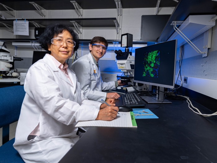
Offering state-of-the-art microscopy instrumentation and services to support our researchers.
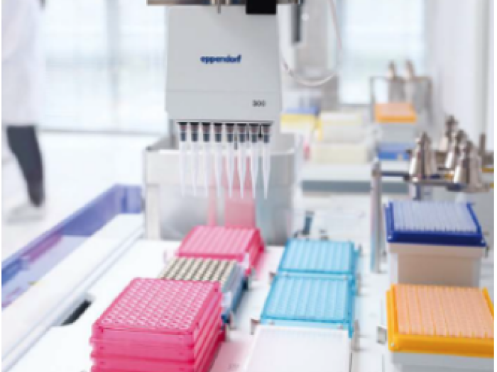
The Microscopy Core offers support and training on a variety of high-end instrumentation and advanced methodologies for both light and electron microscopy, as well as a wide range of sample preparation services for electron microscopy, and training and assistance with image analysis.
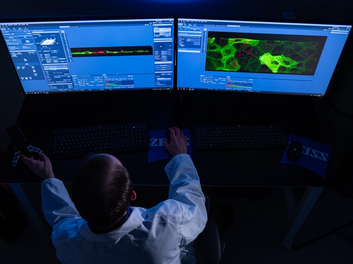
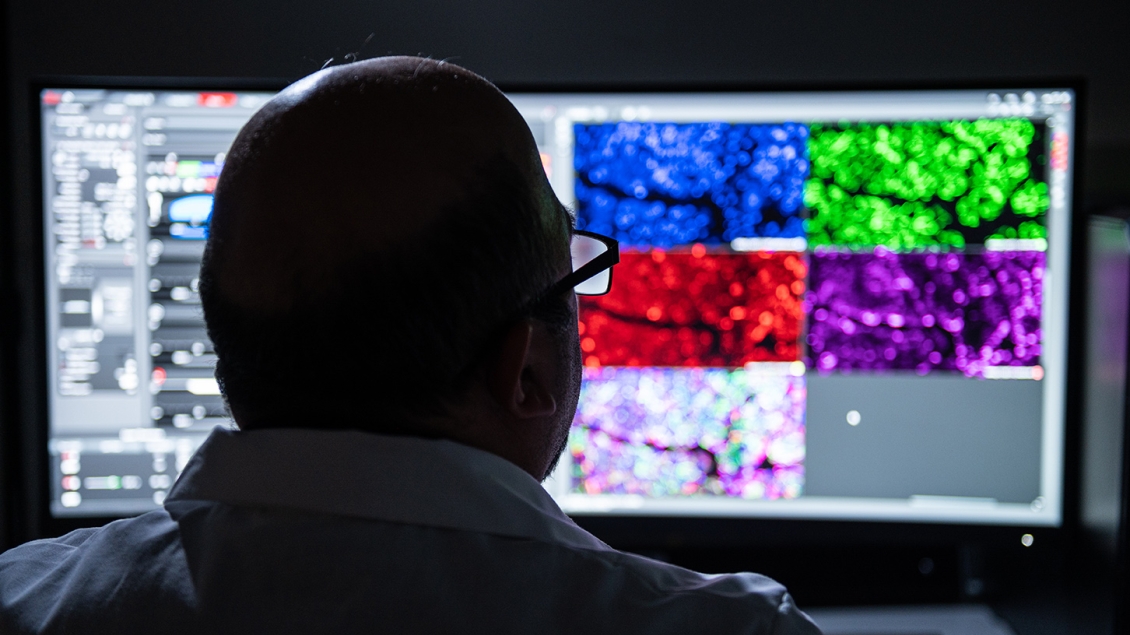
The Microscopy Core houses a wide range of light microscopes and imaging systems. These instruments support a variety of imaging modalities.
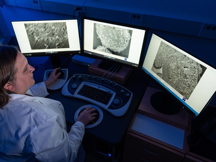
The Microscopy Core owns a basic transmission electron microscope (TEM) and scanning electron microscope (SEM) that are suitable for a large majority of biological EM imaging applications.
In addition to Light Microscopy and Electron Microscopy services, the Microscopy Core provides researchers support across a variety of open-source and commercial image analysis software packages.
Open Source Software:
Commercial Software:
Almost any feature or quality that can be imagined, such as number, size, shape, or intensity, can potentially be measured. Common image analysis requests include: Multi-channel Segmentation, Object Counting, Object Tracking, and 3D Rendering and Visualization.
If you are interested in image analysis, guidance, or education, please contact us.
Rates and fees for routine services are listed below. For complex projects that require extensive labor, please consider including core staff as key personnel on grant submissions. Contact us with any questions you may have.
Rates are subject to change following Office of Financial Analysis review.
| Device | Internal Peak | Internal Off-Peak | Overnight | External |
| JEOL JEM-1400+ LaB6 Transmission Electon Microscope | $40/hr | $30/hr | N/A | $54/hr |
| Leica Stellaris 5 Confocal Microscope | $40/hr | $30/hr | $10/hr | $54/hr |
| Leica SP8 Confocal with 2-Photon or FLIM | $40/hr | $30/hr | $10/hr | $54/hr |
| Leica EM UC7 Ultramicrotome* | $25/hr | $25/hr | N/A | $34/hr |
| Leica Upright SP5 Confocal Microscope with 2-Photon | $40/hr | $30/hr | $10/hr | $54/hr |
| Nikon A1 Confocal | $28/hr | $21/hr | N/A | $38/hr |
| Nikon Ti2 Widefield | $20/hr | $15/hr | $5/hr | $27/hr |
| Nikon N-SIM Super Resolution | $40/hr | $30/hr | $10/hr | $54/hr |
| Nikon X1 Yokogawa Spinning Disk Confocal | $40/hr | $30/hr | $10/hr | $54/hr |
| Retiring Widefields: Nikon E-800 & Zeiss Apotome | $20/hr | $15/hr | N/A | $27/hr |
| Zeiss EVO 15 LaB6 Scanning Electron Microscope | $40/hr | $30/hr | N/A | $54/hr |
| Zeiss 980 LSCM with AiryScan - Installed April 2023 | $40/hr | $30/hr | $10/hr | $54/hr |
| Zeiss Gaussian Lightsheet 7 | $40/hr | $30/hr | $10/hr | $54/hr |
| Zeiss Lattice Lightsheet 7 | $40/hr | $30/hr | $10/hr | $54/hr |
Peak times are Monday - Friday, 8:00 am – 6:00 pm. All other times are Off-Peak. For overnight timelapse experiments over 12 hours only, the time between 12:00 am and 8:00 pm will be charged at the overnight rate. Please inquire regarding the cost and scheduling for timelapse experiments lasting more than 24 hours.
*Skilled users can use an ultramicrotome if they bring their own diamond knife. Training in ultramicrotomy is not typically provided, but could be offered to interested users and would be charged at the labor rate.
| Service | Internal | External |
| Training Per Hour Charge* | $75 | $115 |
| Electron Microscopy Sample Preparation (up to 4 samples) | $400 | $600 |
| Advanced Electron Microscopy Sample Preparation (up to 4 samples) | $950 | $1,425 |
| Ultramicrotome Sectioning: Mesh Grids (per sample) | $150 | $225 |
| Ultramicrotome Sectioning: Slotted Grids (per sample) | $200 | $300 |
| Ultramicrotome Re-sectioning (per block) | $75 | N/A |
| Critical Point Dryer Per Use Charge | $100 | $150 |
| Glow Discharger Per Use Charge | $50 | $75 |
| Additional EM Labor Per Hour Charge* | $75 | $115 |
| Clearing / Expansion Sample Prep (per sample) | $100 | $150 |
| Life Canvas Per Use Charge (excludes reagents) | $50 | $75 |
| Additional LM Labor Per Hour Charge* | $75 | $115 |
*Training is charged at the instrument rate plus the labor rate. Training time is estimated at 2 hours for widefield and EM operation and 3 hours for confocal and advanced light microscopy techniques. These training times are only estimates; more or less time may be required. For the Zeiss LLS7, standard training is not typically offered. A labor charge equivalent to 0.5-1 hour will be added to each experiment for assistance with setup. After several experiments, the user may be permitted to set up their own experiment, at which time the labor charge will be waived. Any service not specified in the rate table, including data analysis services, will be charged at the labor rate.
The Microscopy Core offers resources and workshops to support researchers with image analysis and microscopy. Future events will be added as they are offered. Researchers can also find self-guided resources and presentations from past lectures below.
- Image Analysis Summer Series 2023
- Part I: Basic Optics
- Part II: Trasmitted Techniques
- Part III: Fluorescence Microscopy
- FIJI Quick Start
- FIJI Macro Workshop
- Seeing Double: Key Concepts in Structured Illumination Microscopy
- The Importance of ‘Good’ Probes for High Resolution Imaging
- Confocal and Lattice Light Microscopy
- TIRF and Super Resolution Microscopy
- You Can Observe a Lot by Just Watching!
Biomedical Science Research Building
109 Zina Pitcher Drive, Room A830
North Campus Research Complex
2800 Plymouth Rd, Room 53S
Medical Sciences II
1137 Catherine St, Room 5631
The Microscopy Core is one of the Biomedical Research Core Facilities, and a part of the Medical School Office of Research, where our mission is to foster an environment of innovation and efficiency that serves the Michigan Medicine research community and supports biomedical science from insight to impact.