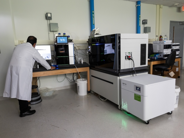
Wide-ranging proteomic services using the latest mass spectrometry technology.
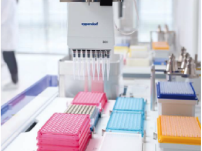
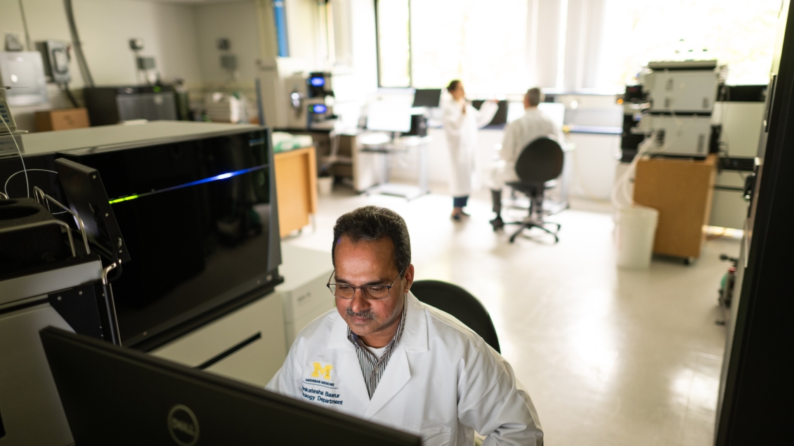
Using state-of-the-art instrumentation, the Proteomics Resource Facility team assists researchers in designing proteomic experiments. We provide technical expertise and bioinformatic support during both discovery and validation phases of proteomic experiments in a cost-effective and timely manner.
We have contributed to over 150 publications, supported numerous successful grant applications, and we are a designated shared resource for the U-M Center for Gastrointestinal Research and the Rogel Cancer Center.
expert identification, analysis, and quantitation of proteins
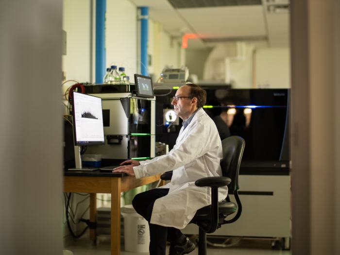
state-of-the-art technology
advanced informatics support
| Service | Fee |
| Protein ID + Fractionation + 90 min LC-MS/MS analysis | $200 |
| Protein ID + Fractionation + 3 hour LC-MS/MS analysis | $319 |
| Relative quantitation (TMT 6-plex) or Phospho-enrichment analysis + Fractionation + 90 min LC-MS/MS analysis | $601+($148/fraction) |
| Relative quantitation (TMT 6-plex) or Phospho-enrichment analysis + Fractionation + 3 hour LC-MS/MS analysis | $601+($267/fraction) |
| Relative quantitation (TMT 10-plex)+ Fractionation + 90 min LC-MS/MS analysis | $1,202+($148/fraction) |
| Relative quantitation (TMT 10-plex)+ Fractionation + 180 min LC-MS/MS analysis | $1,202 +($267/fraction) |
| Relative quantitation (TMT 6-plex) AND Phospho-enrichment analysis + Fractionation + 90 min LC-MS/MS analysis | $1,202+($148/fraction) |
| Relative quantitation (TMT 6-plex) AND Phospho-enrichment analysis + Fractionation + 3 hour LC-MS/MS analysis | $1,202+($267/fraction) |
| Relative quantitation (TMT 10-plex) AND Phospho-enrichment analysis+ Fractionation + 90 min LC-MS/MS analysis | $2,404 +($148/fraction) |
| Relative quantitation (TMT 10-plex) AND Phospho-enrichment analysis+ Fractionation + 3 hour LC-MS/MS analysis | $2,404 +($267/fraction) |
| Service | Fee |
| Parallel Reaction Monitoring (PRM) | Please Inquire |
| Data-Independent Acquisition (DIA) | Please Inquire |
| Service | Fee | Description |
| Protein Identification | $200 | Includes tryptic digestion* + 90 min LC-MS/MS analysis + Database search |
| Relative Quantitation (Ex. TMT)** | $601 | Includes tryptic digestion* + Isotope labeling + 90 min LC-MS/MS analysis + Database search |
| Phospho-Enrichment Analysis** | $601 | Includes tryptic digestion** + Phospho-Enrichment + 90 min LC-MS/MS analysis + Database search |
* Enzymes other than trypsin can be used. User will provide the MS sequencing grade enzymes.
** Quantitation reagent (TMT, or isotope labeled synthetic peptide) and phospho-enrichment kits will be provided by the user.
| Service | Fee/Sample |
| Fractionation followed by 90 min LC-MS/MS analysis | $148 |
| 3 hour LC-MS/MS analysis (Extended gradient) | $119 |
| Funding | Eligibility | Funding or Subsidy Available | Apply |
| U-M Rogel Cancer Center Proteomics Shared Resource | Rogel Cancer Center Members (Core grant) | 30% subsidy on all services; $6,000/PI/year | When submitting samples through iLAB, subsidy is automatically applied to eligible PIs. |
| Proteomics Resource Facility Pilot Project Program | U-M Medical School Faculty | $6,000 (2/3 PRF; 1/3 PI) | TBD |
Please visit iLABs for the most up-to-date services offered and cost information. These rates are reviewed by and approved by the U-M Office of Financial Analysis and are subject to change.
The rates listed are for internal users. For external rates please contact us at [email protected] for a discussion of your project plan and an estimate.
The Proteomics Resource Facility (PRF) uses state-of-the-art, highly sensitive mass spectrometers to fulfill its mission. It is worthwhile to note that while the PRF strives hard to obtain the best results possible, the quality of the data generated by the mass spectrometer is only as good as the quality of the samples introduced into them. Hence it is imperative that the samples intended for MS analysis be prepared by taking extreme care to avoid some of the common contaminants which can impair an otherwise well planned and executed experiment. These contaminants are either “chemical contaminants” or “exogenously introduced protein” contaminants.
Polymers (from plasticware), buffer components (i.e. inorganic salts, detergents), protein stabilizing agents (i.e. glycerol, PEG). While inorganic salts are easily removed by reverse phase-based sample clean-up steps, other contaminants persist even after.
- To minimize polymer contamination, please rinse plastic ware with HPLC grade organic solvent (Methanol or Acetonitrile), if available.
- For biochemical techniques (cell/tissue lysis, IP, affinity purification, etc.) use the least complex buffer required. Some of the commonly used buffers such as RIPA containing small amounts of detergents (ex. 0.1% triton, 0.1% SDS etc) are OK.
- Use the highest quality chemicals/reagents available.
- Please consult with the Laboratory Manager before you begin the experiment to verify the MS compatibility of your buffers components.
Mass spectrometers, under normal operational conditions, are not quantitative (Signal intensity depends on various properties of the peptide/protein including the ionization efficiency). Hence contaminating proteins which may ionize more efficiently and/or co-elute with the peptide of interest can drastically affect the out come of an experiment. Most common contaminants observed are cytokeratin (coming from skin and hair follicles) and serum proteins (i.e. albumin).
- Wear gloves all the time and change them often.
- If possible, keep a small area just for MS sample processing. Wipe the area clean with methanol or ethanol.
- Keep apparatus (staining trays, electrophoresis unit, pipettes, etc.) exclusively for MS sample preparation whenever possible.
- Wash the cells/tissues extensively with isotonic buffer BEFORE lysis (to remove any serum proteins coming from tissue culture medium or blood)
- Use pre-cast gels.
- Handle the gel as little as possible and keep it covered at all times. Consider using StainEase staining trays or equivalent.
- Run a parallel sample for documentation purposes, if possible.
We have experienced satisfactory results with the following protein stains:
- Coomassie stain: Any single-step Coomassie stain that doesn’t require extensive and multiple changes of destaining solution works well. We recommend Colloidal Blue or SimplyBlue stains.
- Silver stain: Classical silver stain techniques that utilize glutaraldehyde and/or formaldehyde are not suitable as they modify amino acid side chains and interfere with digestion with protease. Several MS compatible silver stains are commercially available. We recommend ProteoSilver or SilverQuest kits. Please remember to bring the appropriate destaining solutions when submitting samples.
If you are located on or near the Ann Arbor campus, you can bring the whole gel to our laboratory and we will cut the gel slices for you.
- Please use 1 mm thick gels whenever possible.
- Follow either Coomassie or MS-compatible silver staining protocols.
- Once the band/spot of interest has been located, using a clean scalpel blade, cut the gel as close to the stain as possible.
- If you are planning to process the whole lane, transfer the gel onto a clean glass plate and cut the lane into 12-16 equal sized slices (~0.5 cm each)
- Using a clean pair of forceps, transfer the gel slice(s) into a 1.5 ml eppendorf tube. There is no need to include any buffer/water in the tube.
- This can be kept at -20°C until submission for analysis. If shipping from outside, ship it overnight on dry ice.
- Please ensure that properly filled sample submission forms accompany every sample.
As a general rule, it is safe to assume that many of the commonly used biochemical reagents, especially detergents, may interfere with MS analysis. Please discuss with the Laboratory Director and Manager before preparing the samples.
Please discuss with the Laboratory Director and Manager before preparing the samples.
In most cases, Investigator can follow their optimized protocol for sample preparation. However, after the final wash, we request you to perform 1-2 brief rinses with a simple buffer (ex. PBS or TBS) to remove any residual detergents or protease inhibitors etc. After rinsing, remove all buffer solution and submit just the beads for MS analysis. You may store the beads, after removing buffer solution, at -80 C till you submit.
Other points to consider:
- Use magnetic beads. This format is very useful to remove the final wash buffer almost completely before freezing the beads.
- Use antibody coupled beads, if available.
- If you believe that the final wash with a simple buffer might disrupt your interactome, please discuss with the Lab Manager about the buffer compatibility.
Please discuss with the Laboratory Director and Manager before preparing the samples.
- Use appropriate inhibitors, when available, for sample preparation in order to make sure that the sample is enriched for PTM of interest (ex. phosphtase or proteosome inhbitors).
- As a general rule, PRF will require higher quantities of protein in appropriate buffers for PTM analysis.
Please contact us at 734-615-5722 or [email protected] to discuss your project before preparing samples.
Turn-around time is generally 2 weeks but can vary depending on the complexity and nature of the analyses proposed.
View more publications where the Proteomics Resource Facility has been cited.
| Authors | Year | Publication |
| Schon SB, Yang K, Schindler R, Jiang L, Neff LM, Seeley RJ, Marsh EE. | 2022 | Obesity-related alterations in protein expression in human follicular fluid from women undergoing in vitro fertilization. F S Sci. 2022. PMID: 36096447 |
| Geng X, Wang C, Gao X, Chowdhury P, Weiss J, Villegas JA, Saed B, Perera T, Hu Y, Reneau J, Sverdlov M, Wolfe A, Brown N, Harms P, Bailey NG, Inamdar K, Hristov AC, Tejasvi T, Montes J, Barrionuevo C, Taxa L, Casavilca S, de Pádua Covas Lage JLA, Culler HF, Pereira J, Runge JS, Qin T, Tsoi LC, Hong HS, Zhang L, Lyssiotis CA, Ohe R, Toubai T, Zevallos-Morales A, Murga-Zamalloa C, Wilcox RA | 2022 | GATA-3 is a proto-oncogene in T-cell lymphoproliferative neoplasms. Blood Cancer J. 2022. PMID: 36329027 |
| Bhattacharya A, Wei J, Song W, Gao B, Tian C, Wu SA, Wang J, Chen L, Fang D, Qi L. | 2022 | Sel1L-HRD1 ER-associated degradation suppresses hepatocyte hyperproliferation and liver cancer. iScience 2022:25(10). PMID: 36238898 |
| Kathania M, Kumar R, Lenou ET, Basrur V, Theiss AL, Chernoff J, Venuprasad K. | 2022 | Pak2-mediated phosphorylation promotes ROR_t ubiquitination and inhibits colonic inflammation. Cell Rep. 2022 40(11):111345. doi: 10.1016/j.celrep.2022.111345. PMID: 36103814 |
| Wright SE, Rodriguez CM, Monroe J, Xing J, Krans A, Flores BN, Barsur V, Ivanova MI, Koutmou KS, Barmada SJ, Todd PI. | 2022 | CGG repeats trigger translational frameshifts that generate aggregation-prone chimeric proteins. Nucleic Acids Res. 2022, 50(15):8674-8689. PMID: 35904811 |
| Yang J, Chang Y, Tien JC, Wang Z, Zhou Y, Zhang P, Huang W, Vo J, Apel IJ, Wang C, Zeng VZ, Cheng Y, Li S, Wang GX, Chinnaiyan AM, Ding K. | 2022 | Discovery of a Highly Potent and Selective Dual PROTAC Degrader of CDK12 and CDK13. J Med Chem. 2022 Aug 25;65(16):11066-11083. doi: 10.1021/acs.jmedchem.2c00384. Epub 2022 Aug 8. PMID: 35938508 |
| Abbineni PS, Tang VT, da Veiga Leprevost F, Basrur V, Xiang J, Nesvizhskii AI, Ginsburg D. | 2022 | Identification of secreted proteins by comparison of protein abundance in conditioned media and cell lysates. Anal. Biochem. 2022. PMID: 35973625 |
| Hotta T, McAlear TS, Yue Y, Higaki T, Haynes SE, Nesvizhskii AI, Sept D, Verhey KJ, Bechstedt S, Ohi R. | 2022 | EML2-S constitutes a new class of proteins that recognizes and regulates the dynamics of tyrosinated microtubules. Curr Biol. 2022 Aug 6:S0960-9822(22)01128-9. PMID: 35963242 |
| Herber I, Zedan D, McClintock S, Varani J, Aslam MN. | 2022 | Liver proteomic profile in mice on a high-far diet: Modulation with anti-tumor intervention. FASEB J. 2022. PMID: 35556832 |
| Cerqueira FM, Photenhauer AL, Doden HL, Brown AN, Abdel-Hamid AM, Morais S, Bayer EA, Wawrzak Z, Cann I, Ridlon JM, Hopikins J, Koropatkin NM. | 2022 | Sas20 is a highly flexible starch-binding protein in the Ruminococcus bromii cell-surface amylosome. J. Biol. Chem. 2022. PMID: 35378131 |
Acknowledgment of Contributions
Publications containing data generated by the Proteomics Resource Facility (PRF) should include an acknowledgement of the PRF or include members as coauthors. The PRF uses these acknowledgements and authorship to help demonstrate our contributions to the research community. This in turn helps secure future funding to maintain a robust Core facility and provide professional development of its staff. The Association of Biomolecular Resource Facilities has published a guideline to use when considering whether or not to include core laboratory members on publications.

Professor, Department of Pathology
Professor, Department of Computational Medicine & Bioinformatics
Research Associate Scientist
Department of Pathology
1150 West Medical Center Drive
Ann Arbor, MI 48109